
Skin Cancer on the Face: Early Warning Signs, Treatment Options, and Reconstruction
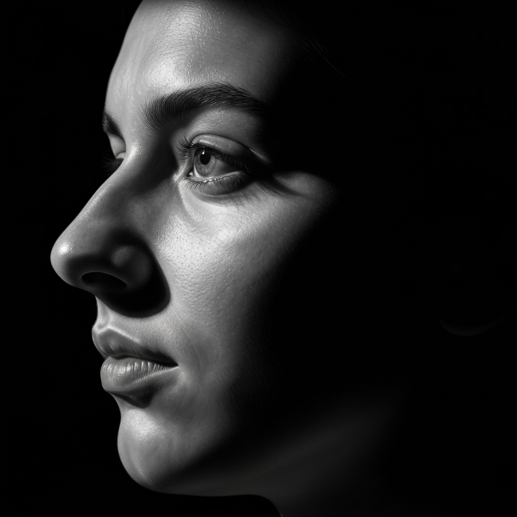
Receiving a diagnosis of skin cancer on the face can be an overwhelming experience. Because the face is our most visible feature, patients naturally have significant concerns about both the complete removal of the cancer and the cosmetic outcome of the treatment. Fortunately, modern medical advancements have made it possible to effectively treat facial skin cancers while simultaneously restoring the natural appearance and function of the affected areas.
Education is the first step in managing skin cancer. This comprehensive guide provides an evidence-based overview of how skin cancer develops, the early warning signs to look for, the diagnostic process, and the advanced treatment and reconstructive options available to patients today.
Understanding Skin Cancer
Skin cancer is the most common type of cancer in the United States. It develops when the DNA within skin cells becomes damaged—most often as a result of cumulative exposure to ultraviolet (UV) radiation from the sun or tanning beds. This damage causes the cells to grow and divide uncontrollably, forming a malignant tumor.
The face, head, and neck are particularly vulnerable to skin cancer because they receive the highest amount of incidental sun exposure throughout a person's lifetime. Because facial skin is relatively thin and sits closely over complex anatomical structures like the nose, eyelids, and lips, early detection and specialized treatment are crucial to ensuring the best possible medical and cosmetic outcomes.
Types of Facial Skin Cancer
There are three primary types of skin cancer that commonly affect the face, head, and neck. Understanding the differences between them can help patients recognize potential issues early.
- Basal Cell Carcinoma (BCC): This is the most common and least dangerous form of skin cancer. BCCs grow very slowly and rarely spread (metastasize) to other parts of the body. However, if left untreated, they can grow deep into surrounding tissues, causing significant local damage.
- Squamous Cell Carcinoma (SCC): SCC is the second most common type of skin cancer. It tends to grow faster than BCC and has a slightly higher risk of spreading to lymph nodes if ignored. SCCs often develop from precancerous lesions called actinic keratoses.
- Melanoma: While less common than BCC and SCC, melanoma is the most serious form of skin cancer. It develops in the melanocytes (the pigment-producing cells) and has a higher tendency to spread to other organs if not caught early.
- Rare Skin Cancers: Less common malignancies, such as Merkel cell carcinoma or dermatofibrosarcoma protuberans (DFSP), can also occur on the face and require specialized multidisciplinary management.
Early Warning Signs
Early detection is the most important factor in successfully treating facial skin cancer. Patients should familiarize themselves with their skin and monitor for any new or changing lesions. Common warning signs include:
- A persistent, non-healing sore that may bleed, ooze, or crust over.
- A pearly, translucent, or shiny bump (often a sign of BCC).
- A red, scaly, or rough patch that may bleed easily (often a sign of SCC).
- A new mole or a change in an existing mole.
- A lesion that grows rapidly or causes localized discomfort.
When evaluating moles for potential melanoma, medical professionals use the ABCDE rule:
- A - Asymmetry: One half of the mole does not match the other half.
- B - Border: The edges are irregular, ragged, or blurred.
- C - Color: The color is not uniform and may include varying shades of brown, black, red, white, or blue.
- D - Diameter: The spot is larger than 6 millimeters (about the size of a pencil eraser), though melanomas can sometimes be smaller.
- E - Evolving: The mole is changing in size, shape, or color.
Risk Factors
While anyone can develop skin cancer, certain factors increase an individual's risk. Understanding these risk factors can help patients take proactive preventative measures.
- Sun Exposure: Cumulative lifetime exposure to UV radiation is the primary cause of most skin cancers.
- Skin Type: Individuals with fair skin, light-colored eyes (blue or green), and blond or red hair are at a higher risk.
- Age: Risk increases with age due to accumulated sun damage.
- Previous Skin Cancer: A history of skin cancer increases the likelihood of developing new lesions.
- Family History: A family history of melanoma or other skin cancers can elevate personal risk.
- Immune Suppression: Patients with weakened immune systems (such as organ transplant recipients) are at significantly higher risk for aggressive skin cancers.
- Indoor Tanning: The use of tanning beds dramatically increases the risk of all types of skin cancer.
How Skin Cancer Is Diagnosed
If you notice a suspicious lesion on your face, it is important to have it evaluated by a dermatologist or a qualified medical specialist. The diagnostic process typically involves:
- Physical Examination: The physician will carefully examine the lesion and your surrounding skin.
- Dermoscopy: A specialized magnifying instrument may be used to evaluate the lesion's pigment patterns and underlying structures.
- Biopsy: The only definitive way to diagnose skin cancer is through a biopsy. A small sample of the suspicious tissue is removed and sent to a pathology laboratory for microscopic analysis.
- Pathology Report: The pathologist will determine if cancer cells are present and identify the specific type and characteristics of the cancer.
- Imaging: In rare cases where a cancer is advanced or suspected to have spread, imaging studies (such as an ultrasound or CT scan) may be ordered.
Treatment Options
Treatment recommendations are highly individualized and depend on the type of cancer, its size, depth, location, and the patient's overall health. Not every suspicious lesion is cancerous, and not every skin cancer requires the same approach. Common treatment options include:
- Observation: In certain low-risk situations or for specific precancerous lesions, close monitoring may be recommended.
- Surgical Excision: The tumor is surgically removed along with a margin of healthy tissue to ensure clear borders.
- Mohs Micrographic Surgery: A specialized, tissue-sparing technique used for high-risk areas like the face.
- Reconstructive Surgery: Procedures performed to repair the defect left after cancer removal.
- Radiation Therapy: May be used as a primary treatment for patients who are not surgical candidates, or as an additional (adjuvant) therapy for high-risk tumors.
- Multidisciplinary Care: Complex cases, such as advanced melanomas, often require collaboration between dermatologists, head and neck surgeons, medical oncologists, and radiation oncologists.
Mohs Surgery Explained
Mohs micrographic surgery is widely used for treating basal cell and squamous cell carcinomas on the face, head, and neck. It is performed by a specially trained dermatologist (a Mohs surgeon).
During the procedure, the visible tumor is removed along with a very thin layer of surrounding tissue. This tissue is immediately examined under a microscope while the patient waits. If cancer cells are seen at the edges, the surgeon removes another thin layer only from the exact area where cancer remains. This process is repeated until the margins are completely clear.
Mohs surgery offers high cure rates for these cancers while preserving as much healthy surrounding tissue as possible—a critical factor when operating on delicate facial areas like the eyelids, nose, or lips. Once the cancer is completely removed, the focus shifts to immediate reconstruction planning.
Facial Reconstruction After Skin Cancer
After a skin cancer is removed from the face, a defect (a "hole" or wound) remains. The goal of facial reconstruction is to repair this defect while restoring both normal function and a natural appearance. Depending on the size and complexity of the defect, the Mohs surgeon may perform the reconstruction, or they may refer the patient to a facial plastic and reconstructive surgeon or head and neck surgeon.
Reconstructive treatment planning is highly individualized. The surgeon carefully evaluates the defect's size, depth, location, and its proximity to critical structures like the eyes, nose, or mouth. Common reconstructive techniques include:
- Primary Closure: The edges of the wound are brought together and sutured in a straight line, often hiding the scar in a natural facial crease.
- Local Flaps: Adjacent healthy skin and tissue are carefully mobilized and shifted to cover the defect. Flaps provide an excellent color and texture match because the tissue comes from the immediate surrounding area.
- Skin Grafts: A piece of skin is taken from another part of the body (such as behind the ear or the collarbone area) and placed over the defect.
- Cartilage Grafting: If structural support is lost (such as on the nose or ear), cartilage may be borrowed from the ear or rib to rebuild the framework.
- Secondary Intention Healing: In some small, shallow defects, the wound may be allowed to heal naturally on its own with meticulous wound care.
The healing process requires patience. Scars will initially appear pink and slightly raised, but they will mature, soften, and fade significantly over the course of 6 to 12 months.
Preventing Future Skin Cancer
Patients who have had one skin cancer are at an increased risk of developing another. Proactive prevention and ongoing surveillance are essential parts of long-term care.
- Daily Sun Protection: Apply a broad-spectrum sunscreen with an SPF of 30 or higher every day, even on cloudy days. Reapply every two hours when outdoors.
- Protective Clothing: Wear wide-brimmed hats, UV-blocking sunglasses, and sun-protective clothing.
- Routine Skin Checks: Schedule regular full-body skin examinations with your dermatologist (typically every 6 to 12 months, as recommended).
- Self-Examinations: Perform monthly self-exams to monitor your skin for any new or changing lesions.
- Avoid Tanning Beds: The use of indoor tanning equipment should be strictly avoided.
- Early Evaluation: If you notice any suspicious changes, do not wait for your next scheduled appointment. Contact your physician promptly for an evaluation.
Common Myths About Skin Cancer
Misinformation can delay diagnosis and treatment. Let's address a few common myths:
- "Only older people get skin cancer." While risk increases with age, melanoma and other skin cancers are increasingly diagnosed in young adults.
- "Skin cancer always hurts." Most skin cancers are completely painless in their early stages. A non-healing sore is a more common warning sign than pain.
- "A mole must change color to be dangerous." While color changes are a warning sign, a mole that changes in size, shape, or begins to bleed or itch should also be evaluated.
- "Sunscreen completely prevents skin cancer." Sunscreen significantly reduces risk, but it must be used in combination with other protective measures, such as seeking shade and wearing protective clothing.
Frequently Asked Questions
What does facial skin cancer look like?
It can vary widely. It may look like a shiny, pearly bump, a red, scaly patch, a non-healing sore, or a new or changing mole. Any persistent, unusual lesion should be evaluated.
Does every skin cancer require Mohs surgery?
No. The treatment depends on the type, size, and location of the cancer. However, Mohs surgery is frequently used for facial cancers due to its tissue-sparing approach.
Will reconstruction leave scars?
Any surgical incision leaves a scar. However, a skilled reconstructive surgeon utilizes advanced techniques to place incisions within natural facial creases and minimize tension, allowing scars to heal as inconspicuously as possible over time.
How is skin cancer diagnosed?
The only definitive way to diagnose skin cancer is through a biopsy, where a small tissue sample is removed and examined under a microscope by a pathologist.
How often should I have skin exams?
Most patients with a history of skin cancer are recommended to have full-body skin exams every 6 to 12 months, though your dermatologist will recommend a schedule based on your specific risk factors.
Can skin cancer return?
Yes. Even with high cure rates, there is a small risk of recurrence. Additionally, patients with a history of skin cancer are at a higher risk of developing new, separate skin cancers, making routine surveillance critical.
Patient Education Resources
We are committed to providing you with the resources necessary to make informed decisions about your care. We encourage you to explore the following areas of our website:
- Mohs Defect Reconstruction
- Skin Cancer Reconstruction
- Facial Reconstruction
- Head & Neck Surgery
- Recovery Guides
- Medical Glossary
- Patient Forms
- Blog
- Areas Served
- Contact
Final Thoughts
A diagnosis of facial skin cancer requires a careful, individualized approach to ensure both complete removal of the disease and the restoration of your natural appearance. By understanding the warning signs, seeking early evaluation, and partnering with experienced specialists for both cancer removal and reconstruction, patients can navigate this process with confidence.
If you have been diagnosed with skin cancer on the face, head, or neck and are seeking reconstructive evaluation, we invite you to contact our office to schedule a comprehensive consultation. Our team is dedicated to providing evidence-based care, transparent education, and compassionate support throughout your treatment and recovery.
Schedule Your Consultation in Metro Detroit
Take the first step towards feeling and looking your best. Contact Dr. Hassan Nasser's office today.
Book an AppointmentRelated Articles
Everything You Need to Know About Thyroid Nodules: Symptoms, Diagnosis, and Treatment
Discover comprehensive, evidence-based information about thyroid nodules, including common symptoms, how they are evaluated with ultrasound and biopsy, and when treatment or surgery is recommended.

How to Choose the Right Facial Plastic Surgeon: 15 Questions Every Patient Should Ask
Choosing a facial plastic surgeon is a significant decision. Learn the 15 essential questions every patient should ask during their consultation to ensure safety, expertise, and natural results.

Mohs Surgery vs. Mohs Reconstruction: Understanding the Difference and Why It Matters
Understand what happens before, during, and after Mohs surgery, and why facial reconstructive surgery is often a critical part of the skin cancer treatment process.